Tuesday, August 20, 2024

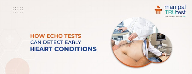
How ECHO Tests Can Detect Early Heart Conditions

Heart disease poses a serious risk to your health. Detecting it early can greatly impact treatment and recovery. One effective tool for early detection is the ECHO test. This simple, painless ultrasound exam produces clear images of your heart. These images help doctors find potential issues before they become severe. It allows for timely intervention and better results.


What is an ECHO Test?

An ECHO test, or echocardiogram, is an ultrasound used to view the heart. It sends sound waves to create live pictures of your heart’s chambers, valves, and blood flow. These images help doctors evaluate how well your heart is working and if there are any issues.

Why Do You Need an ECHO Test?

There are several reasons your doctor might suggest an ECHO test:

       Diagnose Heart Conditions

ECHO tests help identify problems like heart valve disease, heart muscle diseases, and congenital defects.

       Evaluate Heart Function

They show how well your heart pumps blood and reveal any structural abnormalities.

       Monitor Heart Conditions

For those with existing heart issues, regular ECHO tests track disease progress and treatment effectiveness.

How Does an ECHO Test Work?

During an ECHO test, you lie on a table. A technician uses a small device called a transducer on your chest. This device sends sound waves into your heart, creating images. These images appear on a monitor for the doctor to review.

Types of ECHO Tests

Several ECHO tests offer different views of your heart:

     Transthoracic ECHO (TTE): This common type places the transducer on your chest.

     Transesophageal ECHO (TEE): This involves placing the transducer in your esophagus for a closer look.

     Stress ECHO: This test evaluates your heart’s performance during exercise or after medication.

     Doppler ECHO: This measures blood flow through your heart and its chambers.

Preparing for an ECHO Test

Most ECHO tests need little preparation. You can eat and drink as usual. For a stress ECHO, you might need to avoid certain foods or medications. Always inform your doctor about any medications you are taking.

What Happens During an ECHO Test?

An ECHO test is quick and painless, usually lasting 30 to 60 minutes. You lie on an exam table while the technician moves the transducer around your chest. You may need to shift positions to get various views of your heart.

ECHO Test Results

After the test, your doctor will review and explain the images. ECHO results provide details about:

       Heart Size and Shape

Helps assess any enlargements or deformities.

       Heart Muscle Thickness and Movement

Shows how well your heart muscles are functioning.

       Heart Valve Function

Evaluates how well your heart valves are working.

       Blood Flow

Assesses the flow of blood through your heart.

       Abnormal Structures

Identifies any unusual growths or issues.

The Importance of Early Detection

Detecting heart problems early is the main element of effective treatment. ECHO tests are essential in finding heart conditions early, making them easier to manage. Regular checkups and tests like the ECHO can help you keep your heart healthy and address issues before they worsen.

Conclusion

An ECHO test is a valuable, non-invasive way to examine your heart. If your doctor suggests an ECHO test, it's important to proceed without ignoring. A delay may cause your serious side-effects. Early detection of heart issues leads to better treatment options and a higher quality of life. Talk to your doctor about whether an ECHO test is right for you to ensure your heart health is in the best possible condition.
Sunday, August 18, 2024

Understanding PFT (Pulmonary Function Test) and Its Importance

A Pulmonary Function Test (PFT) is a simple exam to check lung health. It helps doctors see how well your lungs work. This test provides clear information on your lung function. Knowing about PFT can help you manage your respiratory health. PFT is vital for detecting lung issues early. It guides treatment and helps in recovery. Understanding PFT's role in lung health is very vital for maintaining good respiratory function. You need to keep track of your lung health with PFT. This ensures timely action if any issues arise.


What is A Pulmonary Function Test?

PFT measures lung performance. It checks how much air you breathe in and out. It also looks at how fast you can breathe. Doctors use special tools for PFT. This test is non-invasive and provides detailed information about lung health.

Why is Pulmonary Function Test Important?

PFT is important for early lung disease detection. Early treatment improves recovery chances. It helps in identifying problems before they become severe. Regular PFT can track treatment progress and adjust plans as needed. This test is a key part of managing and improving lung health.

Who Needs Pulmonary Function Test?

Anyone with breathing issues should consider PFT. Smokers are advised to get regular PFTs. People with known lung conditions should have regular tests. PFT is also useful for those experiencing unexplained coughs or shortness of breath. This test is a proactive step for maintaining lung health.

Types of Pulmonary Function Test

Here are the types of Pulmonary Function Tests:

       Spirometry: Measures how quickly and forcefully you can exhale air.

       Lung Volume Measurement: Determines the total volume of air your lungs can hold.

       Diffusion Capacity Test: Assesses how well oxygen passes from the lungs into the blood.

       Oximetry: Measures the oxygen levels in your blood.

       Peak Flow Meter: Monitors how fast you can blow air out of your lungs.

       Body Plethysmography: Measures lung volume and how much air is left in the lungs after exhaling.

How to Prepare for PFT

Preparing for PFT is quite simple. Inform your doctor about all medications. Avoid smoking before the test. Wear loose, comfortable clothes. Follow any specific instructions given by your doctor for best results. Proper preparation ensures accurate test outcomes.

The PFT Procedure

The PFT procedure is simple and painless. You will sit comfortably and breathe into a tube connected to a machine. The machine records your breathing patterns and lung function. The test usually takes a few minutes. There’s no discomfort involved.

PFT Results

After the test, your doctor will discuss the results. Normal results show healthy lungs. Abnormal results might indicate lung issues. Your doctor will explain what the results mean and suggest any further actions. Understanding your results helps in managing lung health effectively.

PFT and Lung Diseases

PFT is crucial for diagnosing lung diseases. It can detect asthma, COPD, and lung fibrosis. This test helps track disease progression and measure treatment effectiveness. Regular PFT can monitor changes in lung function and adjust treatments as necessary.

Benefits of Pulmonary Function Test

PFT has several benefits. It helps in early detection of lung problems. Early diagnosis leads to better treatment outcomes. PFT provides essential information for managing and treating lung diseases. Regular tests ensure you maintain good lung function over time.

Risks of PFT

PFT is mostly safe. Some people might feel dizzy or lightheaded afterward. These symptoms are usually brief and temporary. There are minimal risks involved, and the benefits of early detection outweigh these minor issues.

Conclusion

PFT is essential for checking and managing lung health. It helps find issues early and guides treatment. Regular PFTs are important for keeping your lungs healthy. If you have lung concerns, talk to your doctor about Pulmonary Function Test. Staying informed and proactive helps protect your lung health and improves your overall well-being. Go for routine health checkup and stay fit and healthy.

Wednesday, August 14, 2024

The Importance of Sonography in Prenatal Care

 From the diagnosis of internal diseases to popularly being used for monitoring the fetus during pregnancy, sonographies are one of the greatest inventions in medical sciences and technology. The Best Women's Health Check-Up Packages may often include a USG scan to check womb health and determine and prevent any threat to the mother or the child during pregnancy.

A USG scan is entirely painless, because it neither requires poking on the skin with sharp objects such as needles or the threats of radiation. Instead, a secure and well-coordinated soundwave is used in the USG device to obtain images of the patientÅ› insides. There are different types of sonography tests used for different purposes and the Best Diagnostic Lab has a clear understanding of what courses to take.


Sonography For Prenatal Care

The ultrasound procedure is highly effective during the prenatal stage allowing accurate visual insight into the womb. From the early stages of the pregnancy, the USG procedure is used for discerning the health of the baby or any complications faced by the mother during this time. Prenatal Ultrasonography is to be conducted from the Best Diagnostic Centre.

A prenatal ultrasound is performed on the mother in different ways to assess the growth of the baby or track any health complications during the pregnancy. The most common type of ultrasound is the Transabdominal Ultrasound which is performed by a professional covering outer skin over the abdominal area of the patient. 

The Best Diagnostic Centre must have the proper technology and equipment to perform a Transvaginal Ultrasound. This procedure is specifically applied for getting clearer pictures of the fetus in the early days of pregnancy. Under this procedure, the vaginal canal of the patient is infiltrated with a transducer to acquire a clear image of the fetus.

Importance Of Ultrasound In Prenatal Care & How To Prepare For It?

Usually, a pregnant woman has to appear for 2-3 ultrasound sessions at the Best Diagnostic Centre, during the 9 months of pregnancy to monitor the health and growth of the baby and ensure fine delivery. The ultrasound scan reference is prescribed by a doctor, once the patient reveals about their pregnancy.

       Ultrasounds are performed on pregnant women most commonly to observe the fetus.

       The scan also provides greater insight into specific medical conditions affecting the pregnancy.

       An Ultrasound from the Best Diagnostic Centre later in the pregnancy can show the prominent growth, movement, heart rate, and bodily features of the baby going as far as revealing a due date. 

       The scan can also be instrumental in the early detection of physical problems with the baby.

As mentioned earlier, Ultrasound is normally harmless and painless but there are other things the patient has to know and follow, for the procedure to produce effective results. There is not much to prepare for an Ultrasound scan at the Best Diagnostic Lab. However, the only thing the patient will be asked to do is drink plenty of water to have ample fluid in their bladder enabling a better view of the insides. If there are other instructions to be followed before the test, your healthcare provider will inform you of the same.

Conclusion

Prenatal care is a vital part of giving birth to a healthy baby and USGs performed at the Best Diagnostic Centre is the first step to ensuring that. Ultrasonography is the most efficient diagnostic tool when it comes to confirming pregnancy and gathering further info leading up to the month of delivery. 

Asthma: Managing Symptoms and Preventing Attacks

Good Health Packages must include tests for Asthma in the early years of life for all, as air pollution and the rise of debilitating lung c...