Heart disease poses a serious risk to your health. Detecting it early can greatly impact treatment and recovery. One effective tool for early detection is the ECHO test. This simple, painless ultrasound exam produces clear images of your heart. These images help doctors find potential issues before they become severe. It allows for timely intervention and better results.
What is an ECHO Test?
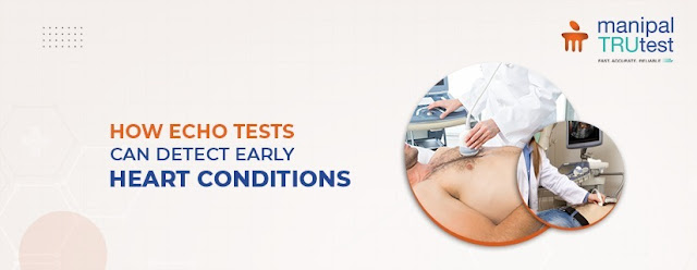
An ECHO test,
or echocardiogram, is an ultrasound used to view the heart. It sends sound
waves to create live pictures of your heart’s chambers, valves, and blood flow.
These images help doctors evaluate how well your heart is working and if there
are any issues.
Why Do You Need an ECHO Test?
There are
several reasons your doctor might suggest an ECHO test:
● Diagnose
Heart Conditions
ECHO tests
help identify problems like heart valve disease, heart muscle diseases, and
congenital defects.
● Evaluate
Heart Function
They show how
well your heart pumps blood and reveal any structural abnormalities.
● Monitor
Heart Conditions
For those
with existing heart issues, regular ECHO tests track disease progress and
treatment effectiveness.
How Does an ECHO Test Work?
During an
ECHO test, you lie on a table. A technician uses a small device called a
transducer on your chest. This device sends sound waves into your heart,
creating images. These images appear on a monitor for the doctor to review.
Types of ECHO Tests
Several ECHO
tests offer different views of your heart:
● Transthoracic
ECHO (TTE): This common
type places the transducer on your chest.
● Transesophageal
ECHO (TEE): This
involves placing the transducer in your esophagus for a closer look.
● Stress
ECHO: This test evaluates your
heart’s performance during exercise or after medication.
● Doppler
ECHO: This measures blood flow
through your heart and its chambers.
Preparing for an ECHO Test
Most ECHO
tests need little preparation. You can eat and drink as usual. For a stress
ECHO, you might need to avoid certain foods or medications. Always inform your
doctor about any medications you are taking.
What Happens During an ECHO Test?
An ECHO test
is quick and painless, usually lasting 30 to 60 minutes. You lie on an exam
table while the technician moves the transducer around your chest. You may need
to shift positions to get various views of your heart.
ECHO Test Results
After the
test, your doctor will review and explain the images. ECHO results provide
details about:
● Heart
Size and Shape
Helps assess
any enlargements or deformities.
● Heart
Muscle Thickness and Movement
Shows how
well your heart muscles are functioning.
● Heart
Valve Function
Evaluates how
well your heart valves are working.
● Blood
Flow
Assesses the
flow of blood through your heart.
● Abnormal
Structures
Identifies
any unusual growths or issues.
The Importance of Early Detection
Detecting
heart problems early is the main element of effective treatment. ECHO tests are
essential in finding heart conditions early, making them easier to manage.
Regular checkups and tests like the ECHO can help you keep your heart healthy
and address issues before they worsen.
Conclusion
An ECHO test
is a valuable, non-invasive way to examine your heart. If your doctor suggests
an ECHO test, it's important to proceed without ignoring. A delay may cause
your serious side-effects. Early detection of heart issues leads to better
treatment options and a higher quality of life. Talk to your doctor about
whether an ECHO test is right for you to ensure your heart health is in the
best possible condition.

.png)


No comments:
Post a Comment